|
|
|


/
 0 0/
/
/


|


- 홈
- > 인체모형
- > 심장 혈관 순환기계
| 상품번호 : 232989 | |||||||
|
|||||||


 |
 (0) (0) |
 (0) (0) |
 |
|---|
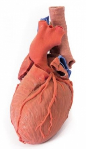
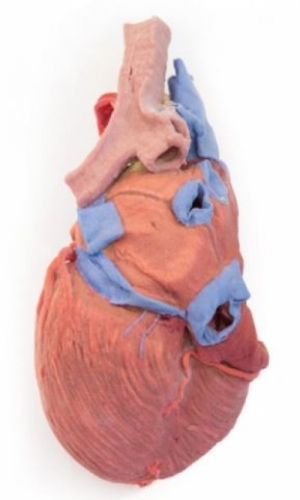
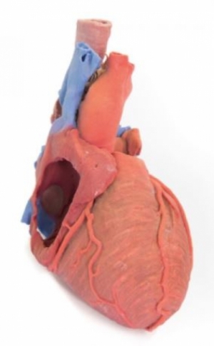
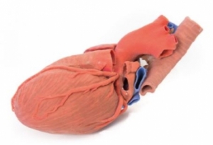
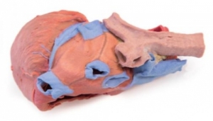
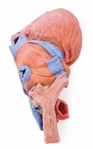
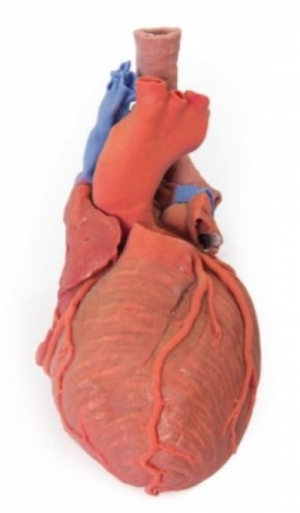
● 이 3D 프린팅 모형은 대혈관과 좌심방(횡심막동 및 사심낭동의 심낭 반사를 보여줌)과 관련된 후방 종격동의    원위 기관, 융기 및 일차 기관지와 심장의 외부 해부학적 구조를 보존합니다.
● 전방 창은 우심방과 귓바퀴 기저부로 절개되어 우방실(삼첨판) 판막과 우심실로 통하는 통로를 노출시킵니다.
● 오른쪽 및 왼쪽 관상동맥과 명명된 가지가 모두 보입니다(후심실간동맥이 오른쪽 관상동맥에서 발생함).
● 왼쪽 귓바퀴는 관상동맥 홈에서 circumflex artery의 경로를 보여주기 위해 절단되었습니다.
● 심장 정맥은 제거되었지만 관상 정맥동은 좌심방 아래로 유지되었습니다.
● 폐동맥을 제거하여 (열린) 폐반월판을 노출시켰으며, 대동맥궁은 손상되지 않아 완두동맥,    왼쪽 총경동맥 및 왼쪽 쇄골하의 기원을 표시합니다.
● 대동맥에 인접하여 좌, 우 상완두 정맥의 종단과 상대정맥으로의 협착 정맥이 보존됩니다.
● This 3D printed specimen preserves the external anatomy of the heart and the distal trachea, carina,    and primary bronchi in the posterior mediastinum relative to the great vessels and left atrium    (which demonstrates the pericardial reflections of the transverse and oblique pericardial sinuses). ● An anterior window has been dissected into the right atrium and base of the auricle, exposing    the right atrioventricular (tricuspid) valve and passage into the right ventricle. ● Both the right and left coronary arteries and named branches are visible    (with the posterior interventricular artery arising from the right coronary artery). ● The left auricle has been sectioned to demonstrate the course of the circumflex artery    in the coronary groove. ● The cardiac veins have been removed, but the coronary sinus has been retained inferior to the left atrium. ● The pulmonary trunk has been removed to expose the (open) pulmonary semilunar valves,    while the arch of the aorta is intact to display the origins of the brachiocephalic trunk,    left common carotid, and left subclavian. ● Adjacent to the aorta, the termination of the left and right brachiocephalic veins and azygos vein    into the superior vena cava is preserved.
 |
(0) |
(0) |
|
|---|
 |
|
|
(0) |
(0) |
|
|---|
 |
|
2. 해외구매 특성상 주문에서 배송까지는 평균 10~15일이 소요됩니다. 간혹 현지 제품 수급에 따라 부득이하게 시일이 더 소요 될 수 있으니 구매시 좀 더 여유있게 주문하시길 권합니다.
3. 해외 내수품인 관계로 A/S에 대해서는 별도의 책임을 지지 않습니다.
4. 해외배송 특성상 주문접수후 배송상태가 배송준비중으로 넘어간 경우 해외에서 국내로의 배송이 이루어지고 있다는 뜻입니다. 따라서 배송준비중으로 배송상태가 넘어간 경우 취소및 반품이 불가하므로 이점 양해 부탁드립니다.
5. 타 해외구매대행 사이트에서 주문하신 물건과 주문날짜가 겹치지않도록 주의해 주십시오. 통관날짜가 같을 경우 합산관세가 부가되게 됩니다.
|


|