|
|
|


/
 0 0/
/
/


|


- 홈
- > 인체모형
- > 심장 혈관 순환기계
| 상품번호 : 232990 | |||||||
|
|||||||


 |
 (0) (0) |
 (0) (0) |
 |
|---|
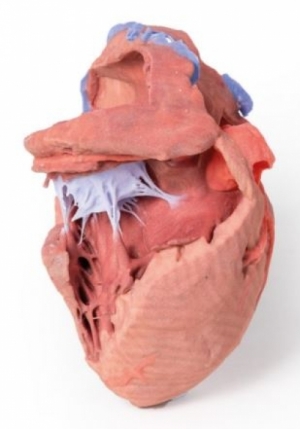
● 이 3D 프린팅 심장 모형은 챔버의 내부 구조를 표시하기 위해 해부되었습니다.
● 심장의 기저부에서는 우심방으로 들어가는 상대정맥의 말단이 보존됩니다.
● 하대정맥의 일부도 우심방의 하측면에 보존되어 있습니다. ● 그러나 대부분의 혈관 내강과 앞쪽 벽의 많은 부분이 제거되어 오른쪽 귓바퀴와    난원와(3D 프린트에서 거의 반투명함)의 pectinate 근육이 노출되었습니다.
● 우심실의 전벽도 제거되어 우심실 판막과 3개의 교두(전방, 후방 및 중격)를 노출시켰습니다.    (심실 중격에서 전방 유두 근육으로 들어가는 것).
● This 3D printed heart has been dissected to display the internal structures of the chambers. ● At the base of the heart the termination of the superior vena cava is preserved entering the right atrium. ● Part of the inferior vena cava is also preserved on the inferior aspect of the right atrium; however,    most of the vessel lumen and much of the anterior wall has been removed to expose    the pectinate muscles of the right auricle and the fossa ovalis (which is nearly translucent in the 3D print). ● The anterior wall of the right ventricle has also been removed to expose the right atrioventricular valve    and its three cusps (anterior, posterior, and septal), including the chordae tendineae connecting them    to respective papillary muscles projecting from trabeculae carneae (including a septomarginal trabecula    entering the anterior papillary muscle from the interventricular septum).
 |
(0) |
(0) |
|
|---|
 |
|
|
(0) |
(0) |
|
|---|
 |
|
2. 해외구매 특성상 주문에서 배송까지는 평균 10~15일이 소요됩니다. 간혹 현지 제품 수급에 따라 부득이하게 시일이 더 소요 될 수 있으니 구매시 좀 더 여유있게 주문하시길 권합니다.
3. 해외 내수품인 관계로 A/S에 대해서는 별도의 책임을 지지 않습니다.
4. 해외배송 특성상 주문접수후 배송상태가 배송준비중으로 넘어간 경우 해외에서 국내로의 배송이 이루어지고 있다는 뜻입니다. 따라서 배송준비중으로 배송상태가 넘어간 경우 취소및 반품이 불가하므로 이점 양해 부탁드립니다.
5. 타 해외구매대행 사이트에서 주문하신 물건과 주문날짜가 겹치지않도록 주의해 주십시오. 통관날짜가 같을 경우 합산관세가 부가되게 됩니다.
|


|